Embark on an in-depth exploration of the phases of mitosis on the whiteboard, an interactive and visually engaging tool that elucidates the intricate process of cell division. This guide will delve into the key stages of mitosis, providing a comprehensive overview and an immersive learning experience.
As cells undergo division, they progress through a series of distinct phases, each characterized by unique events and structural changes. Mitosis, a fundamental process in eukaryotic cells, ensures the faithful distribution of genetic material to daughter cells, maintaining genetic stability and enabling growth and development.
Phases of Mitosis

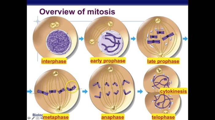
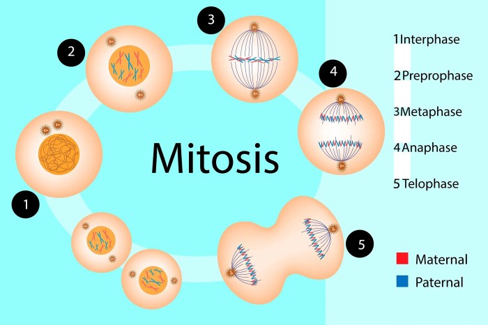
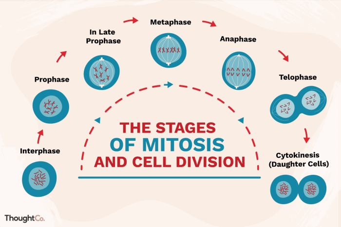
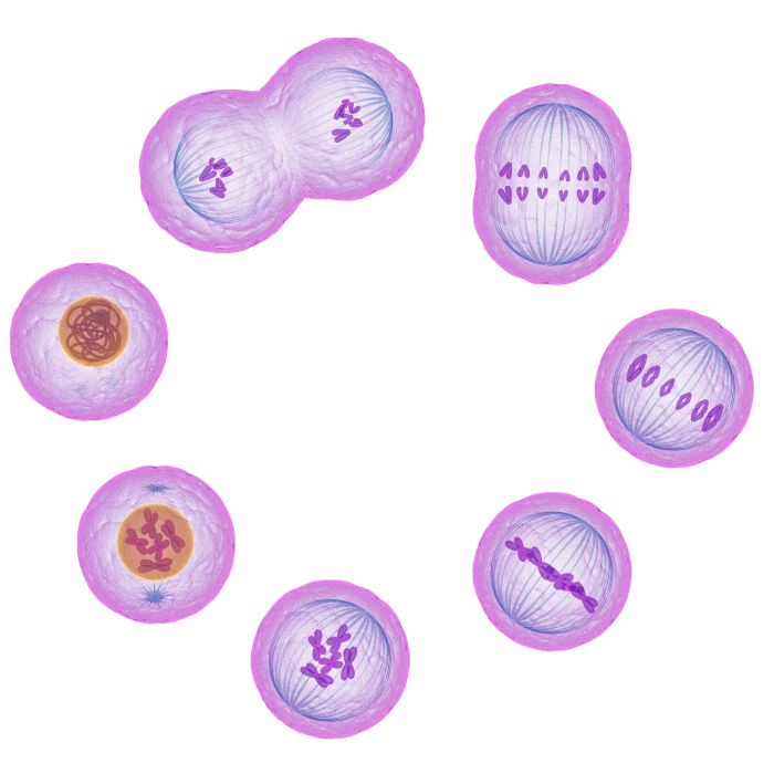
Mitosis is a type of cell division that produces two identical daughter cells from a single parent cell. It is a continuous process, but it can be divided into four distinct phases: prophase, metaphase, anaphase, and telophase.
Phases of Mitosis
| Phase | Description | Image | Key Features |
|---|---|---|---|
| Prophase | The chromosomes become visible and the nuclear envelope breaks down. The centrosomes move to opposite poles of the cell and spindle fibers form between them. | [Image of prophase] |
|
| Metaphase | The chromosomes line up in the center of the cell. The spindle fibers attach to the chromosomes and pull them apart. | [Image of metaphase] |
|
| Anaphase | The chromosomes continue to be pulled apart until they reach opposite poles of the cell. | [Image of anaphase] |
|
| Telophase | Two new nuclear envelopes form around the chromosomes. The spindle fibers disappear and the cell membrane pinches in the middle, dividing the cell into two daughter cells. | [Image of telophase] |
|
Prophase
Prophase marks the beginning of mitosis, characterized by dramatic changes within the cell. During this phase, the chromosomes become visible as distinct, thread-like structures. The nuclear envelope, which encloses the nucleus, gradually disintegrates, allowing the spindle fibers to form and interact with the chromosomes.
Chromosome Condensation, Phases of mitosis on the whiteboard
In prophase, the replicated chromosomes condense and become visible under a microscope. Each chromosome consists of two identical sister chromatids held together by a centromere.
Nuclear Envelope Disintegration
As the chromosomes condense, the nuclear envelope surrounding the nucleus begins to fragment and eventually disintegrates. This breakdown allows the spindle fibers to access and interact with the chromosomes.
Spindle Fiber Formation
During prophase, spindle fibers, composed of microtubules, begin to form in the cytoplasm. These fibers extend from opposite poles of the cell and will play a crucial role in chromosome segregation during anaphase.
Centrosome Separation
In animal cells, the centrosomes, which organize the spindle fibers, separate and migrate to opposite poles of the cell. In plant cells, the spindle fibers form from multiple locations within the cell.
Illustration of Prophase
The following illustration depicts the key events occurring during prophase:
- Condensed Chromosomes:Visible as distinct, thread-like structures.
- Disintegrating Nuclear Envelope:Fragments and eventually breaks down.
- Spindle Fibers:Form and extend from opposite poles of the cell.
- Centrosomes (Animal Cells):Separate and migrate to opposite poles.
Metaphase

Metaphase is the second phase of mitosis, where the chromosomes are aligned at the metaphase plate, an imaginary line that lies across the equator of the cell. This phase is critical for ensuring that each daughter cell receives a complete set of chromosomes during cell division.
Alignment of Chromosomes
During metaphase, the mitotic spindles, composed of microtubules, attach to the centromeres of each chromosome. The centromere is a specialized region of the chromosome that connects the two sister chromatids, which are identical copies of the original chromosome. The microtubules from opposite poles of the spindle pull on the centromeres, aligning the chromosomes at the metaphase plate.
The alignment of chromosomes at the metaphase plate ensures that each daughter cell receives one copy of each chromosome. This is essential for maintaining the correct number of chromosomes in each daughter cell and preventing genetic abnormalities.
Diagram of Chromosome Arrangement
The following diagram illustrates the arrangement of chromosomes at the metaphase plate:
|
   V
   ^
    \   /
     \ /
      V

-----
  /     \
 /       \
|         |
In this diagram, the metaphase plate is represented by the horizontal line, and the chromosomes are shown as vertical lines.
The centromeres of the chromosomes are attached to the spindle fibers, which are represented by the diagonal lines. The microtubules from opposite poles of the spindle pull on the centromeres, aligning the chromosomes at the metaphase plate.
Anaphase

Anaphase is the third phase of mitosis. During anaphase, the sister chromatids of each chromosome separate and move to opposite poles of the cell.
The separation of sister chromatids is driven by the shortening of microtubules from the mitotic spindle. As the microtubules shorten, they pull the sister chromatids apart. The shortening of microtubules is driven by the motor protein dynein.
Movement of Chromosomes
As the sister chromatids separate, they move towards opposite poles of the cell. This movement is driven by the motor protein kinesin. Kinesin binds to the microtubules and moves along them, pulling the chromosomes behind it.
The movement of chromosomes towards opposite poles of the cell continues until all of the chromosomes have reached their respective poles. Once all of the chromosomes have reached their poles, anaphase is complete and telophase begins.
Telophase: Phases Of Mitosis On The Whiteboard

Telophase is the final stage of mitosis, during which the chromosomes reach the poles of the cell and the nuclear envelope reforms around each set of chromosomes. This results in the formation of two daughter cells, each with its own nucleus and a complete set of chromosomes.
The following events occur during telophase:
- The chromosomes continue to move towards the poles of the cell.
- The nuclear envelope reforms around each set of chromosomes.
- The mitotic spindle fibers disappear.
- The cytoplasm divides into two parts, resulting in the formation of two daughter cells.
Differences between Mitosis and Meiosis
Mitosis and meiosis are two distinct types of cell division that occur in eukaryotic cells. Mitosis produces two daughter cells that are genetically identical to the parent cell, while meiosis produces four daughter cells that are genetically different from the parent cell.
| Feature | Mitosis | Meiosis |
|---|---|---|
| Number of daughter cells | 2 | 4 |
| Genetic identity of daughter cells | Identical to parent cell | Different from parent cell |
| Number of cell divisions | 1 | 2 |
| Synapsis | No | Yes |
| Crossing over | No | Yes |
FAQ Resource
What is the significance of prophase in mitosis?
Prophase marks the initiation of mitosis, characterized by chromatin condensation and the formation of visible chromosomes. During this phase, the nuclear envelope breaks down, and spindle fibers begin to assemble, preparing the cell for chromosome segregation.
How do chromosomes align during metaphase?
In metaphase, chromosomes align at the metaphase plate, a central plane within the cell. This alignment ensures that each chromosome is positioned correctly for separation during anaphase.
What is the role of sister chromatids in anaphase?
Anaphase is characterized by the separation of sister chromatids, which are identical copies of each chromosome. These chromatids are pulled apart by spindle fibers, moving towards opposite poles of the cell.